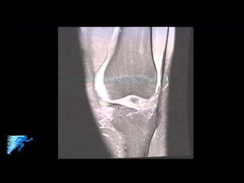
Deep Medial Capsular Ligament Knee
The static stabilizers of the medial knee include the superficial MCL s-MCL the deep MCL d-MCL or medial capsular ligament and the posterior oblique ligament 5 10. The medial tibial collateral ligament MCL of the knee is a flat triangular band on its medial aspect and has superficial and deep portions.
 What To Do About Meniscal Injuries Exercises For Injuries Knee Joint Anatomy Knee Problem Knee Joint
The Deep medial ligament dMCL is divided into two the meniscofemoral and meniscotibial ligaments.

Deep medial capsular ligament knee. Its connected below to the medial condyle of the tibia above the groove for the tendon of semimembranosus. The deep MCL can be divided into 2 parts. The knee is the joint most commonly examined at magnetic resonance MR imaging.
Ligaments of Knee Joint. Deep fascia of the lower extremity to cover the gastrocne-mius and popliteal fossa. It has two components.
Patients who underwent ACL reconstruction for complete. Layer III is the deep layer including the joint capsule and d-MCL. And 2 the meniscotibial or coronary ligament Figure 7.
It is more common in the medial more frequently posterior horn region 5 than in the lateral compartment of the knee. The origin of the meniscofemoral comes from the femur just distal to the superficial medial collateral inserting into the medial menisci. Layer 1 deep fascia.
View larger version 91K Fig. The purpose of this study was to investigate the incidence and patterns of medial collateral ligament complex injuries in patients with clinically isolated ACL ruptures. Medial ligaments and capsule are primary and secondary stabilizers of valgus rotation and anterior and posterior translation.
Clinical findings are nonspecific and can include pain instability and joint effusion. A joint recess that is in continuity with the intraarticular joint space is located posterior to the medial femoral condyle and deep in relation to the capsule and medial gastrocnemius tendon. Also called the deep medial ligament or middle capsular ligament.
Anterior third middle third and posterior third Fig. Its primary function is to resist outward turning forces on the knee. 1 the meniscofemoral ligament.
The supporting structures on the medial side of the knee consist of a superficial fascial layer I a deep capsular layer III with the deep medial collateral ligament in it and in between the superficial collateral ligament layer II. Layer 2 superficial Medial Collateral Ligament MCL layer 3 joint capsule and the deep MCL. The s-MCL is a broad ligament that attaches at the medial femoral epicondyle and inserts just below the pes.
The deep medial capsular ligament is a thickening of the medial joint capsule which is most distinct anteriorly where it parallels the TCL. The superficial portion of the MCL contributes 57 and 78 of medial stability at 5 degrees and 25 degrees of knee flexion respectively. The medial collateral ligament MCL or tibial collateral ligament TCL is one of the four major ligaments of the knee.
Superiorly the knee capsule joins the medial gastrocnemius tendon. Layer II intermediate comprises the s-MCL and medial patellofemoral ligament MPFL. The medial and posteromedial regions of the knee are important for knee stability but also frequently injured.
This complex is the major stabilizer of the medial knee. The deep part of the ligament is short and mixes together with the fibrous capsule and with the peripheral margin of the medial meniscus. Alternatively the medial side of the knee can be divided into thirds.
It is on the medial inner side of the knee joint in humans and other primates. Existing classifications of MCL lesions do not address involvement of the superficial and deep portions of the MCL or involvement of the anterior vertical and posterior oblique portions. Of these two ligaments the meniscotibial ligament is shorter and thicker.
Although the medial collateral ligament MCL is frequently injured descriptions of the appearance of the medial capsular and supporting structures of the knee at MR imaging are often not very detailed 1. In anterior cruciate ligament ACL injuries concomitant damage to peripheral soft tissues is associated with increased rotatory instability of the knee. Introduction The posteromedial corner of the knee PMC is comprised of the structures between the posterior border of the superficial medial collateral ligament SMCL and the medial border of the posterior cruciate ligament PCL.
The meniscotibial ligament is thicker and shorter. The anatomy of the medial side of the knee has been described by two different approaches layered approach. The meniscotibial coronary ligament and meniscofemoral ligament which attach to the tibia and femur respectively.
The medial knee was divided into the static and dynamic stabilizers. Ramp lesions are a specific type of meniscocapsular injury associated with ACL-deficient knees 6. These ligaments have also been called the medial collateral ligament MCL tibial collateral ligament mid-third capsular ligament and oblique fibers of the sMCL respectively.
The medial ligament complex of the knee is composed of the superficial medial collateral ligament sMCL deep medial collateral ligament dMCL and the posterior oblique ligament POL. In the setting of rupture of the cruciate ligaments it is important to identify injuries in this region because it can possibly alter the treatment strategy and even delay or prevent successful reconstruction of the cruciate ligaments. Tibial Collateral Ligament Fibular Lateral Collateral Ligament.
The anterior third consists of capsular ligaments covered by the extensor retinaculum of the quadriceps. The superficial MCL is the primary stabilizer to valgus stress at all angles deep portion medial capsular ligament separated from superficial portion by a bursa.
 Acl Rupture And Associated Structural Injuries Of The Knee Acl Recovery Acl Rupture Knee Surgery
 Knee Joint Shoulder Anatomy Human Knee Muscle Diagram
 Leg And Knee Anatomy Knee Joint Joints Anatomy Anterior Leg Muscles
 Mcl Tear Of The Right Knee Ligament Tear Mcl Injury Knee Joint Anatomy
 Tim Jul A Medial Meniscus Tear Is More Common Than A Lateral Meniscus Tear Because It Is Firmly Attached To The Meniscus Tear Knee Mri Medial Meniscus Tear
 Knee Interior Views Anatomy Patellar Ligament Medial Patellar Retinaculum Blended Into Joint Capsule Suprapatellar Synov Anatomy Cruciate Ligament Capsule
 Viewing Playlist Aa Junepin Radiopaedia Org Radiology Nuclear Medicine Osteopathy
 Ligaments Anatomy Poster Ligaments Anatomical Chart Company Joints Anatomy Physical Therapy Massage Therapy
 Medial Collateral Ligament Of The Knee Everything You Need To Know Dr Nabil Ebraheim Youtube Knee Ligaments Meniscal Tear Orthopedic Surgery
 Human Knee Anatomy Knee Joint Anatomy Anatomy Of The Knee Bursitis Knee
 Shoulder Sergery Shoulder Anatomy Upper Limb Anatomy Shoulder Joint Anatomy
 Djon Djon Human Anatomy And Physiology Knee Muscles Anatomy Muscle Anatomy
 Patellar Fracture Of Left Knee Medical Malpractice Cases Knee Trials
 Knee Tendonitis The Five Main Areas Around The Knee Joint That Are Typically Afflicted With Tendonitis Knee Tendonitis Knee Injury Treatment Knee Arthritis
 Type Of Joint Found In The Knee And Elbow Koibana Info Synovial Joint Knee Joint Anatomy Anatomy Organs
 9 18 Knee Joint Human Anatomy Model Medical Anatomy Body Anatomy


