Posteromedial Capsule Knee
The medial and posteromedial regions of the knee are important for knee stability but also frequently injured. It also acts as a secondary external rotation stabilizer.
 Approach To Posteromedial Meniscocapsular Junction In Anterior Cruciate Download Scientific Diagram
The post is hinged allowing it to be dropped for lateral and posteromedial arthroscopic instrumentation.
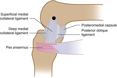
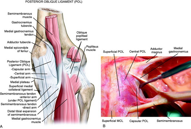
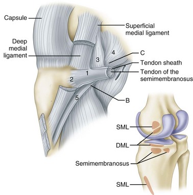
Posteromedial capsule knee. The POL therefore is not a stand-alone structure but a thickening of the posteromedial joint capsule. The post is placed lateral to the surgical knee and 4 inches above the superior pole of the patella. The posterior oblique ligament POL is the main component of the posteromedial corner PMC of the knee and plays a crucial role in acting as a secondary restraint against translation rotation and valgus forces.
Clinical image of the skin incision for the postero-medial approach. Posteromedial capsular releases and the concurrent postoperative rehabilitation program were effective in the treatment of knee extension deficits. With the knee in slight flexion make a straight or slightly curved incision running from the medial epicondyle towards the posteromedial edge of the tibia.
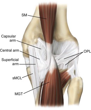
SM The semimembranosus tendon is a part of the posteromedial joint capsule which is important in controlling anteromedial rotatory instability. Posterior Capsulotomy of the Knee A 2-cm incision is then performed on the posteromedial capsule with an electrocautery to arrive at a full exposure of the posterior compartment. Capsule c is seen along posteromedial and posterolateral sides and is inseparable from synovial layer.
Intervention to the posteromedial capsule is performed in the appropriate patient based on physical exam preoperative imaging and arthroscopy as an additional procedure to ACLR Table 1. Provides access to posteromedial side of joint posterior aspect of the medial meniscus posteromedial corner incise the fascia along the anterior border of sartorius retract the sartorius posteriorly together with semitendinosis and gracilis. We believe that arthro-scopic posteromedial capsular releases are an effective.
The encouraging results of this study compare well to data presented in open posterior capsular release studies. The bursae of the knee are synovial-lined sacs that decrease the friction of moving structures. Pain and swelling of the bursa are caused by inflammation external pressure or overuse.
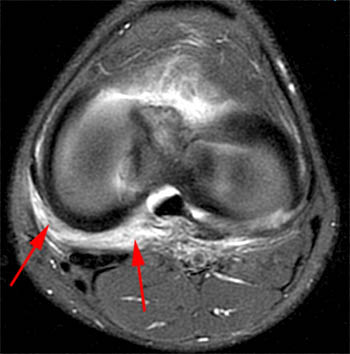
It is used to place valgus stress on the knee opening the medial compartment for arthroscopic access. Capsule is discontinuous at intercondylar area arrow. The posteromedial corner of the knee PMC is an important anatomic structure that is easily seen but often overlooked on magnetic resonance MR images.
In complex knee injuries the patient is positioned supine with both knees in extension. Injury to one of the main structures comprising the posteromedial corner PMC of the knee with modern MRI systems the major anatomic structures comprising the PMC can be readily identified these structures contribute to the static and dynamic stability of the knee including a supporting role in multiligament knee injuries. Arthroscopic posteromedial capsular releases of the knee can result in improved knee motion postoperatively.
Note subgastrocnemius bursa b and Bakers cyst B on posteromedial aspect of knee. Medial ligaments and capsule are primary and secondary stabilizers of valgus rotation and anterior and posterior translation. The posterior oblique ligament POL belongs to the medial supporting structures of the knee and is one of the five components of the posteromedial corner PMC of the knee.
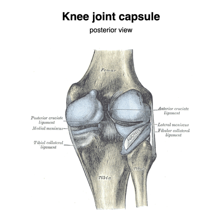
Posteromedial Joint Capsule and Posterior Horn of the Medial Meniscus The knee joint capsule part of layer III forms the deep MCL with its meniscotibial and meniscofemoral components along the medial aspect of the knee. The incision can be extended as needed both proximally and distally as indicated by the dashed line. At intercondylar area synovium covers cruciate ligaments and adjacent fat stars.
It stabilizes internal rotation of the knee through all degrees of flexion but bears the most load when internally rotated in full extension. Importantly it can result in increased stress on the cruciate ligaments and can result in anteromedial rotatory instability AMRI of the knee. The posteromedial corner of the knee PMC is comprised of the structures between the posterior border of the superficial medial collateral ligament SMCL and the medial border of the posterior cruciate ligament PCL.
Together with the pes anserinus tendons it gives medial and posteromedial reinforcement. An arthroscopic posteromedial capsular release involves sectioning the posteromedial capsule at its meniscofemoral portion midway between its femoral attachment and its posterior horn medial meniscus junction. Posteromedial corner injury of the knee is a readily identifiable but frequently underappreciated injury on imaging.
 Medial Ligamentous Injuries Of The Knee Acute And Chronic Musculoskeletal Key
 Medial And Anterior Knee Anatomy Clinical Gate
![]() Illustrations Of A Medial Meniscal Allograft Transplantation For A Download Scientific Diagram
Illustrations Of A Medial Meniscal Allograft Transplantation For A Download Scientific Diagram
 Medial View Of A Right Knee Illustrating The Medial Structures The Download Scientific Diagram
 Medial And Posteromedial Ligament Injuries Diagnosis Operative Techniques And Clinical Outcomes Clinical Gate
Musculoskeletal Key
 Knee Capsule Radiology Reference Article Radiopaedia Org
 Knee Injuries Musculoskeletal Key
.jpg) Posteromedial Corner Injury Knee Sports Orthobullets
Posteromedial Corner Injury Knee Sports Orthobullets
 Pdf Surgical Approaches To The Posteromedial And Posterolateral Aspects Of The Knee Semantic Scholar
 Posterolateral And Posteromedial Corner Injuries Of The Knee Radiology Key
 The Knee Clinical Gate
Https Pubs Rsna Org Doi Pdf 10 1148 Rg 2015140166
 Inside Out Meniscus Repair A Review Asian Journal Of Arthroscopy
 Demystifying The Posteromedial Corner Of The Knee Nyu Langone Orthopedic Digital Library
 Posteromedial Corner Injuries Of The Knee Clinical Radiology
 Posteromedial Corner Injury Of The Knee Radsource
 Posteromedial Approach A For Focused Exposure A 3 Cm Vertical Download Scientific Diagram
 Exploration Of The Posteromedial Compartment Right Knee The Scope Is Download Scientific Diagram